

Testicular Cancer Stages
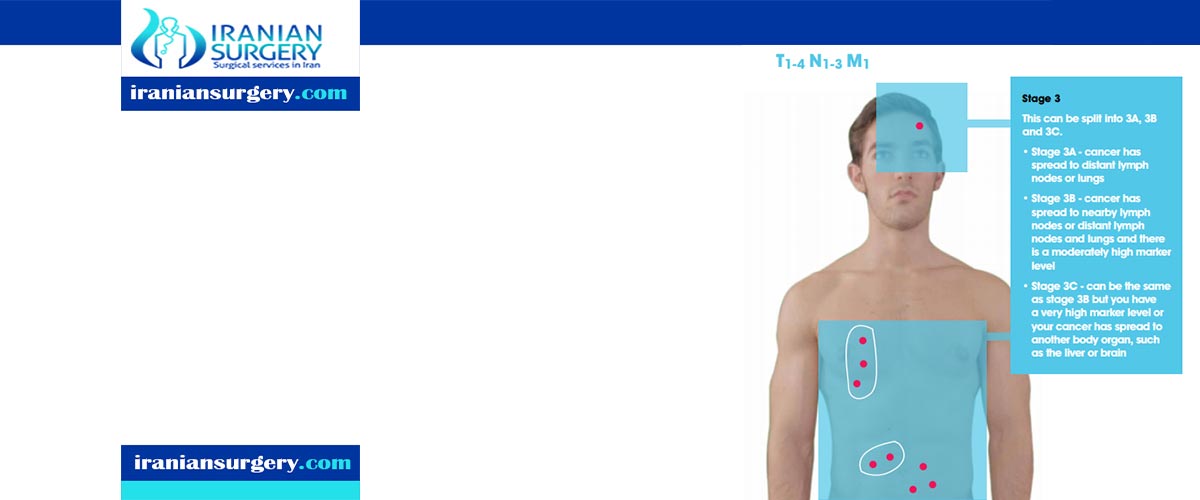
Testicular Cancer Stages
Is Stage 3 testicular cancer curable?
Staging is a way of describing if and where a cancer has spread. Doctors use diagnostic tests, including CT scans and blood tests, to find out the cancer's stage, so staging may not be complete until all of the tests are finished. Knowing the stage helps the doctor to decide what kind of treatment is best and helps predict a patient's prognosis, which is the chance of recovery. There are different stage descriptions for different types of cancer.
Read more about : What size of ovarian cyst is dangerous?
Read more about : Virgin tightening surgery before and after photo
Read more about : Sign of Anal cancer
About Iranian Surgery
Iranian surgery is an online medical tourism platform where you can find the best Surgeons to treat your Testicular cancer in Iran. The price of treating a Testicular cancer in Iran can vary according to each individual’s case and will be determined by the type of treatment you have and an in-person assessment with the doctor. So if you are looking for the cost of Testicular cancer treatment in Iran, you can contact us and get free consultation from Iranian surgery.

TNM staging system
One tool that doctors use to describe the stage is the TNM system. For testicular cancer, an S is added to the TNM system. Doctors use the results from diagnostic tests and scans to answer these questions:
. Tumor (T): How large is the primary tumor? Where is it located?
. Node (N): Has the tumor spread to the lymph nodes in the back of the abdomen (retroperitoneum)?
. Metastasis (M): Has the cancer spread to other parts of the body? If so, where and how much?
. Serum tumor marker (S): Are the serum tumor markers AFP, beta-hCG, and LDH elevated? If so, how high are they?
The results are combined to determine the stage of cancer for each person. There are 3 stages of testicular cancer: stages I, II, and III (1, 2, and 3). The stage provides a common way of describing how advanced the cancer is so that doctors can work together to plan the best treatment. Stage I is called the least advanced or earlier stage, and stage III is called the most advanced or later stage. Patients with the least advanced stages are more likely to be cured and often need less aggressive treatment than patients with a more advanced stage.
Staging for testicular cancer can also be clinical or pathological:
. Clinical staging is based on the results of tests done before surgery, which may include physical examinations and imaging tests. For example, clinical stage II testicular cancer means that the retroperitoneal lymph nodes are enlarged when viewed with a CT or MRI scan.
. Pathological staging is based on what is found during surgery. For example, pathological stage II testicular cancer means that cancer has been found when tissue removed from the retroperitoneal lymph nodes is examined under a microscope. In general, pathological staging provides the most information to determine a patient’s prognosis, but it is not always needed.
Read more about : Foods that cure fissures
Read more about : sleeping position for fissure
Read more about : How to burst a bartholin cyst at home?
Read more about side effects of having only one testicle
Here are more details on each part of the TNM system for testicular cancer:
Tumor (T)
Using the TNM system, the "T" plus a letter or number (0 to 4) is used to describe the size and location of the tumor. Tumor size is measured in centimeters (cm). A centimeter is roughly equal to the width of a standard pen or pencil.
Stage may also be divided into smaller groups that help describe the tumor in even more detail. For testicular cancer, the T stage can only be determined when tissue removed during surgery is examined under a microscope. This means that the T stage is only determined after the testicle is removed, and the T stage is always a pathological stage and never a clinical stage. The “p” before the T stage indicates that it is a pathological stage. Specific tumor stage information is below.
. pTX: The primary tumor cannot be evaluated. If a man has not had the testicle(s) surgically removed, the term "TX" is used.
. pT0 (T plus zero): There is no evidence of a primary tumor in the testicles.
. pTis: This stage describes germ cell neoplasia in situ (GCNIS). This is a precancerous condition in which there are germ cells that appear cancerous but are not yet behaving the way cancer cells do. GCNIS becomes cancer when the cells grow into parts of the testicle(s) where they do not normally belong.
. pT1: The primary tumor is only in the testicle, which may include the rete testis. It has not grown into blood vessels or lymph vessels in the testicles. The tumor may have grown into the inner membrane layer surrounding the testicle, called the tunica albuginea. It has not spread to the outer membrane layer surrounding the testicle, called the tunica vaginalis.
For a pure seminoma, this stage is further divided based on the side of the tumor:
. pT1a. The tumor is smaller than 3 centimeters (cm) in size.
. pT1b. The tumor is 3 cm or larger in size.
. pT2: The tumor is in the testicle, which may include the rete testis, and it has grown into 1 or more of the following parts of the testicle:
. Blood vessels or lymphatic vessels in the testicle
. The epididymis,
. The fatty tissue next to the epididymis called the hilar soft tissue
. The tunica vaginalis
. pT3: The tumor has grown into the spermatic cord.
. pT4: The tumor has grown into the scrotum.
Read more about 2nd iui success rate
Read more about Virgin tightening surgery before and after
Read more about Ovarian cyst size chart
Read more about Cancer treatment in Iran
Read more about rectal bleeding
Read more about Laser Eye Surgery in Iran
Node (N)
The “N” in the TNM staging system stands for lymph nodes. These tiny, bean-shaped organs help fight infection. Lymph is a fluid that flows from the different tissues and organs of the body and eventually drains into the blood stream. It passes through specialized tubes called lymphatic vessels and is filtered along the way by the lymph nodes. Cancer cells often build up and grow in lymph nodes before they spread to other parts of the body. The first place the lymphatic fluid from the testicles drains to is the retroperitoneal lymph nodes located in the back of the abdomen in front of the spine, an area called the retroperitoneum. These are called the regional lymph nodes for testicular cancer. Lymph nodes in the pelvis, chest, or other parts of the body are called distant lymph nodes, even though the testicles are closer to the pelvis than to the retroperitoneum.
In men with testicular cancer, lymph nodes usually are not biopsied or removed. Instead, the N stage (lymph node stage) is most often estimated by using CT scans. N stage that is based on CT scans is the clinical stage. When the N stage is based on a biopsy or removal of the lymph nodes, it is the pathological stage. When a stage has been determined pathologically, the letter “p” is added as the first letter of the stage (for example pN1). The letter "c" stands for clinical stage.
. NX: The regional lymph nodes cannot be evaluated.
. cN0: There is no spread to regional lymph nodes as seen on imaging tests.
. pN0: There is no cancer found in lymph nodes removed during a retroperitoneal lymph node dissection.
. cN1: Imaging tests show signs that the cancer has spread to 1 or more lymph nodes in the retroperitoneum. None of the lymph nodes are bigger than 2 centimeters (cm).
. pN1: There is cancer in 1 to 5 lymph nodes, and none are larger than 2 cm.
. cN2: Imaging tests show at least 1 enlarged lymph node or lymph node mass in the retroperitoneum that is larger than 2 cm but not larger than 5 cm.
. pN2: Either or both of the following conditions:
. There is cancer in more than 5 lymph nodes, but none are larger than 5 cm.
. There is cancer in at least 1 lymph node, and the largest lymph node or lymph node mass is between 2 cm and 5 cm in size.
. cN3: Imaging tests show at least 1 enlarged lymph node or a lymph node mass in the retroperitoneum larger than 5 cm.
. pN3: There is cancer in at least 1 enlarged lymph node or lymph node mass that is larger than 5 cm.
Metastasis (M)
The "M" in the TNM system describes whether the cancer has spread to other parts of the body, called distant metastasis. When testicular cancer spreads, it most commonly spreads to the lung and the lymph nodes of the chest, pelvis, and the base of the neck. More advanced stages may have spread to the liver and bones. Testicular cancer rarely spreads to the brain unless the primary tumor is a choriocarcinoma.
. MX: Distant metastasis cannot be evaluated.
. M0: The disease has not metastasized to distant lymph nodes or other organs.
. M1: There is at least 1 distant metastasis.
. M1a: There is cancer in the lungs or lymph nodes other than the retroperitoneal lymph nodes.
. M1b: The cancer has spread to organs other than a lung. The lungs may or may not also be involved. For example, a testicular cancer that has spread to the liver or the bones is stage M1b.
Serum tumor markers (S)
Serum tumor markers also help to stage testicular cancer. Blood tests for tumor markers will be done before and after surgical removal of the testicle(s). Tumor marker levels usually decrease after the surgery. Generally, the levels need to be tested until they stop decreasing or begin to rise to determine the correct "S" stage. For patients who will receive chemotherapy, the tumor marker levels on the first day of chemotherapy are used to determine the patient’s risk group (see below).
. SX: Tumor marker levels are not available, or the tests have not been done.
. S0: Tumor marker levels are normal.
. S1: At least 1 tumor marker level is above normal. LDH is less than 1.5 times the upper limit of the normal range, beta-hCG is less than 5,000 mIu/mL, and/or AFP is less than 1,000 ng/mL.
. S2: At least 1 tumor marker level is substantially above normal. This means that LDH is 1.5 to 10 times the upper limit of the normal range, beta-hCG is 5,000 to 50,000 mIu/mL, and/or AFP is 1,000 to 10,000 ng/mL. None of the tumor markers is elevated high enough to qualify as S3 (see below).
. S3: At least 1 or more tumor marker level is very highly elevated. This means that LDH is more than 10 times the upper limit of the normal range, beta-hCG is more than 50,000 mIu/mL, and/or AFP is more than 10,000 ng/mL.
Cancer stage grouping
Doctors assign the stage of the cancer by combining the T, N, and M classifications and the S level information.
. Stage 0: Refers to carcinoma in situ, also called intratubular germ cell neoplasia (pTis, N0, M0, S0).
. Stage I: Cancer is at any T level, and there is no evidence of spread to either lymph nodes or other organs. Serum tumor marker levels have not been done or are not available (any T, N0, M0, SX).
. Stage IA: The cancer is only in the testicle. It may have grown into the rete testis, but it has not grown into the epididymis, hilar soft tissue, or lymphatic or blood vessels in the testis. It has not spread to lymph nodes or distant sites. The tumor in the testis may have grown into the inner membrane surrounding the testis, called the tunica albuginea, but not the outer membrane, called the tunica vaginalis. If the tumor is pure seminoma, it is smaller than 3 cm. Serum markers are normal (pT1, N0, M0, S0).
. Stage IB: The testicular tumor has grown into the epididymis, hilar soft tissue, tunica vaginalis, the blood or lymphatic vessels within the testicle, the spermatic cord, or the scrotum. The cancer has not spread to lymph nodes or distant sites. Serum markers are normal (pT2, pT3, or pT4, and N0, M0, S0).
. Stage IS: Cancer is of any T stage and has not spread to lymph nodes or distant sites. Serum markers remain higher than normal levels after the cancerous testicle has been removed (any T, N0, M0, and S1-S3). Stage IS non-seminoma testicular cancer is treated the same as stage III testicular cancer.
. Stage II: The cancer has spread to any number of regional lymph nodes but not to lymph nodes in other parts of the body or distant organs. Serum markers are unavailable (any T, N1-N3, M0, SX).
. Stage IIA: Cancer has spread to retroperitoneal lymph nodes, either clinical or pathological stage N1, but none are larger than 2 cm. If a lymph node dissection has been done, no more than 5 lymph nodes contain cancer. In addition, serum tumor marker levels are normal or only slightly high. There are no signs of cancer having spread anywhere other than the retroperitoneum (any T, N1, M0, S0 or S1).
. Stage IIB: Cancer has spread to lymph nodes in the retroperitoneum, and the largest lymph node with cancer or lymph node mass is between 2 cm and 5 cm in size. If a lymph node dissection has been done, cancer has spread to at least 1 lymph node (or lymph node mass) between 2 cm and 5 cm or to more than 5 lymph nodes, with none larger than 5 cm. Serum marker levels are normal or slightly high. There is no evidence of cancer having spread anywhere other than the retroperitoneum (any T, N2, M0, S0 or S1).
. Stage IIC: Cancer has spread to at least 1 lymph node (or lymph node mass) that is larger than 5 cm. Serum marker levels are normal or slightly high. There is no evidence of cancer having spread anywhere other than the retroperitoneum (any T, N3, M0, S0 or S1).
. Stage III: Cancer has spread to distant lymph nodes or to any organ. Serum tumor marker levels are unknown (any T, any N, M1, SX).
. Stage IIIA: Cancer has spread to distant lymph nodes and/or the lungs. Serum marker levels are normal or only mildly increased (any T, any N, M1a, S0 or S1).
. Stage IIIB: Cancer has spread to any lymph nodes and/or the lungs but not to any other organs. At least 1 serum marker is substantially elevated (any T, N1-N3, M0, S2; or any T, any N, M1a, S2).
. Stage IIIC: Either or both of the following:
. At least 1 serum marker is extremely high, and the cancer has spread to at least 1 lymph node or organ (any T, N1-N3, M0, S3; or any T, any N, M1a, S3).
. The cancer has spread to an organ other than the lungs (any T, any N, M1b, and any S).
Recurrent: Recurrent cancer is cancer that has come back after treatment. If the cancer does return, there will be another round of tests to learn about the extent of the recurrence. These tests and scans are often similar to those done at the time of the original diagnosis.
Later-stage testicular cancer: risk group classification
If the disease has spread to lymph nodes or other organs, the following system is used to classify a germ cell tumor into a good-risk, intermediate-risk, or poor-risk group. This helps to determine the treatment plan and the likelihood of cure. Patients with a tumor in the intermediate and poor-risk groups usually receive more chemotherapy than patients with a tumor in the good-risk category.
The differences between good-risk, intermediate risk and poor-risk are the same as the differences between stage IIIA, stage IIIB, and stage IIIC (above). Stage IIIA is the same as good risk, IIIB is the same as intermediate risk, and IIIC is the same as poor risk.
Good risk
. Non-seminoma. The cancer has not spread to an organ other than the lungs and serum tumor marker levels are good, which means:
AFP less than 1,000 ng/mL
B-hCG less than 5,000 iU/L
LDH less than 1.5 x ULN
. Seminoma. The cancer has not spread to an organ other than the lungs and AFP, any B-hCG, any LDH levels are normal.
Intermediate risk
. Non-seminoma. The cancer has not spread to an organ other than the lungs and the serum tumor marker levels are intermediate, which means:
AFP between 1,000 and 10,000 ng/mL
B-hCG between 5,000 and 50,000 iU/L
LDH between 1.5 x ULN and 10 x ULN
. Seminoma. The cancer has spread to an organ other than the lungs and AFP, any B-hCG, any LDH levels are normal.
Poor risk
. Non-seminoma. The cancer has spread to an organ other than the lungs or the serum tumor marker levels are poor, which means:
AFP is 10,000 ng/mL or higher
B-hCG is 50,000 iU/L or higher
LDH is 10 x ULN or higher
. Seminoma. There are no patients with poor-risk seminoma
10 common questions about Testicular Cancer Stages
[kkstarratings]