

Breast lump biopsy
breast lump biopsy
A biopsy is one of the several tests you may have to check for breast cancer if you have a breast lump. It involves taking a sample of cells or tissue from the area of your breast that needs to be checked. For many biopsies, you'll get an injection to numb the area of the breast to be biopsied.
Types of breast biopsy include:
- Fine-needle aspiration biopsy. This is the simplest type of breast biopsy and may be used to evaluate a lump that can be felt during a clinical breast exam. For the procedure, you lie on a table. While steadying the lump with one hand, your doctor uses the other hand to direct a very thin needle into the lump.

The needle is attached to a syringe that can collect a sample of cells or fluid from the lump. Fine-needle aspiration is a quick way to distinguish between a fluid-filled cyst and a solid mass and, possibly, to avoid a more invasive biopsy procedure. If, however, the mass is solid, a tissue sample will be obtained.
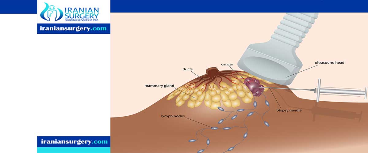
- Core needle biopsy. This type of breast biopsy may be used to assess a breast lump that's visible on a mammogram or ultrasound or that your doctor feels (palpates) during a clinical breast exam. A radiologist or surgeon uses a thin, hollow needle to remove tissue samples from the breast mass, most often using ultrasound guidance.
Several samples, each about the size of a grain of rice, are collected and analyzed to identify features indicating the presence of disease. Depending on the location of the mass, other imaging techniques, such as a mammogram or MRI, may be used to guide the positioning of the needle to obtain the tissue sample.
- Stereotactic biopsy. This type of biopsy uses mammograms to pinpoint the location of suspicious areas within the breast. For this procedure, you generally lie face down on a padded biopsy table with one of your breasts positioned in a hole in the table, or you may have the procedure in a seated position. You may need to remain in this position for 30 minutes to one hour.
The table is raised several feet, and the equipment used by the radiologist is positioned beneath the table. Your breast is firmly compressed between two plates while mammograms are taken to show the radiologist the exact location of the area for biopsy.
The radiologist makes a small incision — about 1/4-inch long (about 6 millimeters) — into your breast. He or she then inserts either a needle or a vacuum-powered probe and removes several samples of tissue. The samples are sent to a lab for analysis.
- Ultrasound-guided core needle biopsy. This type of core needle biopsy involves ultrasound — an imaging method that uses high-frequency sound waves to produce precise images of structures within your body. During this procedure, you lie on your back or side on an ultrasound table.
Holding the ultrasound device (transducer) against your breast, the radiologist locates the mass within your breast, makes a small incision to insert the needle and takes several core samples of tissue to be sent to a lab for analysis.
- MRI-guided core needle biopsy. This type of core needle biopsy is done under guidance of an MRI — an imaging technique that captures multiple cross-sectional images of your breast and combines them, using a computer, to generate detailed 3-D pictures. During this procedure, you lie face down on a padded scanning table. Your breasts fit into a hollow depression in the table.
The MRI machine provides images that help determine the exact location for the biopsy. A small incision about 1/4-inch long (about 6 millimeters) is made to allow the core needle to be inserted. Several samples of tissue are taken and sent to a lab for analysis.
At the time of the breast biopsy procedures noted above, a tiny stainless-steel marker or clip may be placed in your breast at the biopsy site. This is done so that if your biopsy shows cancer cells or precancerous cells, your doctor or surgeon can locate the biopsy area to remove more breast tissue surgically (known as the surgical biopsy).
- Surgical biopsy. During a surgical biopsy, a portion of the breast mass is removed for examination (incisional biopsy) or the entire breast mass may be removed (excisional biopsy, wide local excision or lumpectomy). A surgical biopsy is usually done in an operating room using sedation given through a vein in your hand or arm (intravenously) and a local anesthetic to numb your breast.
If the breast mass can't be felt, your radiologist may use a technique called wire localization to map the route to the mass for the surgeon. During wire localization, the tip of a thin wire is positioned within the breast mass or just through it. This is usually done right before surgery.
During surgery, the surgeon will attempt to remove the entire breast mass along with the wire. To help ensure that the entire mass has been removed, the tissue is sent to the hospital lab to confirm whether breast cancer has been detected and if so, the edges (margins) of the mass are evaluated to determine whether cancer cells are present in the margins (positive margins).
If cancer cells are present at the margins, you will be scheduled for another surgery so more tissue can be removed. If the margins are clear (negative margins), then the cancer has been removed adequately.
10 common questions about Breast lump biopsy
[kkstarratings]