

Skull Base Surgery
Skull Base Surgery
Skull base surgery may be done to remove both noncancerous and cancerous growths, and abnormalities on the underside of the brain, the skull base, or the top few vertebrae of the spinal column. Because this is such a difficult area to see and reach, skull base surgery may be done by a minimally invasive endoscopic procedure. In this procedure, the surgeon inserts instruments through the natural openings in the skull—the nose or mouth—or by making a small hole just above the eyebrow. This type of surgery requires a team of specialists that may include ENT (ear, nose, and throat) surgeons, maxillofacial surgeons, neurosurgeons, and radiologists.
Before endoscopic skull base surgery was developed, the only way to remove growths in this area of the body was by making an opening in the skull. In some cases, this type of surgery may be needed.

What is skull base surgery used for?
These are some of the growths and conditions that may be treated by skull base surgery:
- Cysts that develop from birth
- Growths caused by infections
- Pituitary tumors
- Meningiomas, noncancerous tumors that grow from the meninges, the tissue that covers the brain and lies between the brain and skull
- Chordomas, a slow-growing bone tumor most often found at the base of the skull
- Trigeminal neuralgia, an intense pain on one side of the face
- Craniopharyngiomas, growths that occur near the pituitary gland
- Craniosynostosis, a condition in which the bones of the skull of an infant close too early, causing problems with brain growth and the shape of the skull
- Cerebrospinal fluid fistulas
- Cerebral aneurysm, a weak, often bulging area in a blood vessel in the brain
- Arteriovenous malformations, arteries and veins that are abnormally connected to one another
Types of skull base surgery
Skull base surgery can be done in two main ways. Although the preferred method is endoscopic, open surgery is also an option, depending on the type of growth that needs to be removed and its location:
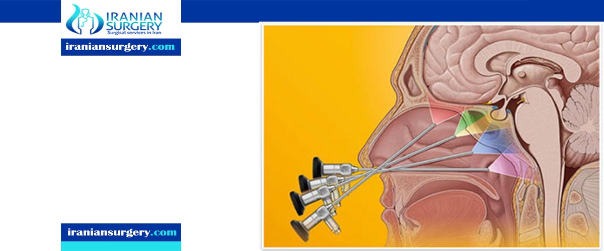
- Endoscopic or minimally-invasive skull base surgery. This type of surgery usually does not require a large incision. A surgeon may make a small opening inside the nose to allow a neurosurgeon to remove a growth through a thin lighted tube called an endoscope. An MRI is a type of picture taken of the skull base using magnets and a computer and may be done by a radiology specialist while the surgical specialists are operating to help them make sure all of the growth has been removed.
- Traditional or open skull base surgery. This type of surgery may require incisions in the facial area and in the skull. Parts of bone may need to be removed so that the growth can be reached and removed. An operating room microscope is often used for this type of surgery.
Symptoms
You may have many possible symptoms from a growth or abnormality in the skull base area. Symptoms will depend on the size, type, and location of the growth or abnormality, and may include:
- Facial pain
- Headache
- Dizziness
- Visual problems
- Numbness
- Weakness of the face
- Hearing loss or ringing in the ears
- Nasal congestion or frequent sinus infections
Diagnosis
The diagnosis of growths or abnormalities that may require skull base surgery is based on your symptoms and a physical exam. Because this area can't be seen directly, these exams and imaging studies are important parts of the diagnosis:
- Brain imaging studies. Special tests such as MRI (magnetic resonance imaging), MRA (magnetic resonance angiogram), PET (positron emission tomography) and CT (computed tomography) scans create pictures of the skull to help your medical team see a growth or abnormality.
- Biopsy. A small piece of a growth in the skull base may be taken out and looked at under a microscope. A biopsy may be done using an endoscope placed through the nose and sinuses. Biopsies may also be done by fine need aspiration, or excisional biopsy.
- Other tests. Your balance, cranial nerves, muscle activity, vision, and hearing may all be checked. Studies or scans of other areas and systems of the body may also be checked.
Treatment
In addition to endoscopic and open skull base surgery, these treatments may be needed, depending on the type of growth or abnormality of the skull base:
- Chemotherapy. These are drugs used to treat growths caused by cancer.
- Radiation therapy. X-ray treatment may be used to control a growth in the skull base that can't be completely removed by surgery.
- Gamma knife. This is a special type of radiation therapy that uses precise X-ray beams to target a growth in the skull base.
- Proton beam therapy. This is another type of radiation therapy designed to have greater accuracy and dosing for tumors.
- Particle therapy. This is the newest form of radiotherapy. It uses high energy particles with fewer side effects. Carbon-ion radiotherapy is a form of particle therapy.
Managing after skull base surgery
After skull base surgery, you will be closely cared for by your medical team. Their goal is to allow you to return to normal activities of daily living. Some people need continued therapy, including radiotherapy, radiosurgery, and proton beam therapy. Many will need repeated imaging to make sure that a growth is not coming back over time. Because this type of surgery can be very stressful, it's also important to get support from friends and family. Your treatment team can also provide mental health and support group resources for you and your family.
10 common question about Skull Base Surgery
[kkstarratings]
1 Comment
We welcome you all across the world to attend the 5th International conference on Gastroenterology and Hepatology which is going to be held on June 29-30, 2020 at Rome, Italy which includes prompt keynote presentations, Poster presentations, oral talks and Exhibitions. We are cordially inviting all Clinical & Medical Experts who are interested in sharing their research experience and practical knowledge in the arena of clinical and medical science.
Theme of the Conference is “Recent Advancements in Gastroenterology and Hepatology”
For more details regarding Scientific Tracks, Submission of Abstract and Registration Please go through: https://gastroenterology.alliedacademies.com/