

Glial Tumor
Glioma
What is a Glioma?
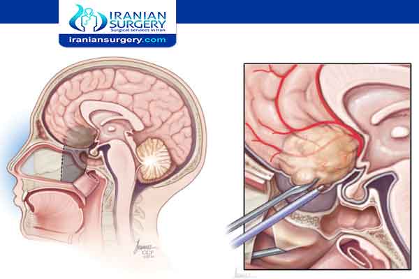
Glioma is a type of tumor that occurs in the brain and spinal cord. Gliomas begin in the gluey supportive cells (glial cells) that surround nerve cells and help them function. Gliomas are one of the most common types of primary brain tumors.
Gliomas are classified according to the type of glial cell involved in the tumor, as well as the tumor's genetic features, which can help predict how the tumor will behave over time and the treatments most likely to work.
A glioma can affect your brain function and be life-threatening depending on its location and rate of growth.
Read more about : Brain cancer treatment
Read more about Do arachnoid cysts require surgery?
About Iranian Surgery
Iranian surgery is an online medical tourism platform where you can find the best Surgeons to treat your Brain Tumor in Iran. The price of treating a Brain Tumor in Iran can vary according to each individual’s case and will be determined by the type of Brain Tumor treatment you undergo and an in-person assessment with the doctor. So if you are looking for the cost of Glioma treatment in Iran, you can contact us and get free consultation from Iranian surgery.

Before Glioma Treatment
Symptoms
The symptoms of glioma vary by tumor type as well as the tumor's size, location and rate of growth.
Common signs and symptoms of gliomas include:
. Headache
. Nausea or vomiting
. Confusion or a decline in brain function
. Memory loss
. Personality changes or irritability
. Difficulty with balance
. Urinary incontinence
. Vision problems, such as blurred vision, double vision or loss of peripheral vision.
. Speech difficulties
. Seizures, especially in someone without a history of seizures
When to see a doctor
Make an appointment with your doctor if you have any signs and symptoms common to glioma.
Causes
Like most primary brain tumors, the exact cause of gliomas is not known. But there are some factors that may increase your risk of a brain tumor.
Read more about Ovarian cyst size chart
Risk factors
Like most primary brain tumors, the exact cause of gliomas is not known. But there are some factors that may increase your risk of a brain tumor. Risk factors include:
. Your age. Your risk of a brain tumor increases as you age. Gliomas are most common in adults between ages 45 and 65 years old. However, a brain tumor can occur at any age. Certain types of gliomas, such as ependymomas and pilocytic astrocytomas, are more common in children and young adults.
. Exposure to radiation. People who have been exposed to a type of radiation called ionizing radiation have an increased risk of brain tumor. Examples of ionizing radiation include radiation therapy used to treat cancer and radiation exposure caused by atomic bombs.
More-common forms of radiation, such as electromagnetic fields from power lines and radiofrequency radiation from microwave ovens have not been shown to increase the risk of glioma.
It isn't clear whether cellphone use increases the risk of brain cancer. Some studies have found a possible association between cellphone use and a type of brain cancer called acoustic neuroma. Many other studies have found no association. Because cellphones are a relatively new factor, more long-term research is needed to understand the potential impact on cancer risk. For the time being, if you're concerned about the possible link between cellphones and cancer, experts recommend limiting your exposure by using a speaker or hands-free device, which keeps the cellphone itself away from your head.
. Family history of glioma. It's rare for glioma to run in families. But having a family history of glioma can double the risk of developing it. Some genes have been weakly associated with glioma, but more study is needed to confirm a link between these genetic variations and brain tumors.
Read more about : Arachnoid cyst size chart
Read more about: benign spinal tumor treatment
Diagnosis
If your primary care doctor suspects you have a brain tumor, you may be referred to a specialist who is trained in treating brain and nervous system disorders (neurologist). Your doctor may recommend a number of tests and procedures, including:
. A neurological exam. During a neurological exam, your doctor may check your vision, hearing, balance, coordination, strength and reflexes. Problems in one or more of these areas may provide clues about the part of your brain that could be affected by a brain tumor.
. Imaging tests. Magnetic resonance imaging (MRI) is often used to help diagnose brain tumors. In some cases, a dye (contrast material) may be injected through a vein in your arm during your MRI study to help show differences in brain tissue.
A number of specialized MRI scan components — including functional MRI, perfusion MRI and magnetic resonance spectroscopy — may help your doctor evaluate the tumor and plan treatment.
Other imaging tests may include computerized tomography (CT) scan and positron emission tomography (PET).
. Tests to find cancer in other parts of your body. To rule out other types of brain tumors that may have spread from other parts of the body, your doctor may recommend tests and procedures to determine where the cancer originated. Gliomas originate within the brain and are not the result of cancer that has spread (metastasized) from elsewhere.
. Collecting and testing a sample of abnormal tissue (biopsy). Depending on the location of the glioma, a biopsy may be performed with a needle before treatment or as part of an operation to remove the brain tumor.
A stereotactic needle biopsy may be done for gliomas in hard-to-reach areas or very sensitive areas within your brain that might be damaged by a more extensive operation. During a stereotactic needle biopsy, your neurosurgeon drills a small hole into your skull. A thin needle is then inserted through the hole. Tissue is removed through the needle, which is frequently guided by CT or MRI scanning.
The biopsy sample is then analyzed under a microscope to determine if it's cancerous or benign.
A biopsy is the only way to definitively diagnose a brain tumor and give a prognosis to guide treatment decisions. Based on this information, a doctor who specializes in diagnosing cancer and other tissue abnormalities (pathologist) can determine the grade or stage of a brain tumor.
The pathologist will also examine the physical appearance and growth rate of your biopsy sample (molecular diagnosis). Your doctor will explain the pathologist's findings to you. This information helps guide decision-making about your treatment plan.
During Glioma Treatment
What are the different types of glioma?
. Astrocytomas are glial cell tumors developed from connective tissue cells called astrocytes and are the most common primary intra-axial brain tumor, accounting for nearly half of all primary brain tumors. They are most often found in the cerebrum (the large, outer part of the brain), but also in the cerebellum (located at the base of the brain).
Astrocytomas can develop in adults or in children. High-grade astrocytomas, called glioblastoma multiforme, are the most malignant of all brain tumors. Glioblastoma symptoms are often the same as those of other gliomas. Pilocytic astrocytomas are low-grade cerebellum gliomas commonly found in children. In adults, astrocytomas are more common in the cerebrum.
. Brain stem gliomas, also called diffuse infiltrating brainstem gliomas, or DIPGs, are rare tumors found in the brain stem. They usually cannot be surgically removed because of their remote location, where they intertwine with normal brain tissue and affect the delicate and complex functions this area controls. These tumors occur most often in school-age children where they are responsible for the greatest number of childhood deaths from primary brain tumors.
. Ependymomas develop from ependymal cells lining of the ventricles or in the spinal cord. Ependymomas are rare, accounting for just 2 percent to 3 percent of primary brain tumors. However, they account for about 8 percent to 10 percent of brain tumors in children, and are more likely to affect those younger than 10 years old. The most location for ependymomas in children is near the cerebellum, where the tumor can block the flow of the cerebral spinal fluid and cause increased pressure inside the skull (obstructive hydrocephalus.) These tumors can spread to other parts of the brain or spinal cord (drop-metastases) due to the flow of spinal fluid.
. Mixed gliomas (also called oligo-astrocytomas) are made up of more than one type of glial cell. Their diagnosis as a distinct tumor type is controversial and may be resolved with genetic screening of tumor tissue. These tumors are often found in the cerebrum and are most common in adult men.
. Oligodendrogliomas form from oliogodendrocytes, the supportive tissue cells of the brain and are usually found in the cerebrum. About 2 percent to 4 percent of primary brain tumors are oliogodendrogliomas. They are most common in young and middle-aged adults and more likely to occur in men. Seizures are a very common symptom of these gliomas (affecting 50 percent to 80 percent of patients), as well as headache, weakness, or problems with speech. Oligodendrogliomas typically have a better prognosis than most other gliomas.
. Optic pathway gliomas are a type of low-grade tumor found in the optic nerve or chiasm, where they often infiltrate the optic nerves, which send messages from the eyes to the brain. People with neurofibromatosis are more likely to develop them. Optic nerve gliomas can cause vision loss and hormone problems, since these tumors are often located at the base of the brain where hormonal control is located. Gliomas affecting hormone function may be known as hypothalamic gliomas.
Treatment
Treatment for glioma depends on the type, size, grade and location of the tumor, as well as your age, overall health and preferences.
In addition to actions to remove the tumor itself, treatment for glioma may also require using drugs to reduce the signs and symptoms of your tumor.
Your doctor may prescribe steroids to reduce swelling and relieve pressure on affected areas of the brain. Anti-epileptic drugs may be used to control seizures.
Read more about : Chemotherapy
Glioma treatment options
. Surgery
Surgery to remove as much of the tumor as possible is usually the first step in treating most types of gliomas.
In some cases, gliomas are small and easy to separate from surrounding healthy brain tissue, which makes complete surgical removal possible. In other cases, tumors can't be separated from surrounding tissue, or they're located near sensitive areas in your brain and make surgery risky. In these situations your doctor removes as much of the tumor as is safe.
Even removing a portion of the tumor may help reduce your signs and symptoms.
In some cases, neuropathologists may analyze tissue samples removed by a surgeon and report the results while surgery is underway. This information helps the surgeon decide how much tissue to remove.
A variety of surgical technologies and techniques may be used to assist the neurosurgeon in protecting as much healthy brain tissue as possible while removing the tumor, including computer-assisted brain surgery, intraoperative MRI, awake brain surgery and lasers. For example, during awake brain surgery, you may be asked to perform a task with the goal of ensuring the area of the brain controlling that function is not damaged.
Surgery to remove a glioma carries risks, such as infection and bleeding. Other risks may depend on the part of your brain in which your tumor is located. For instance, surgery on a tumor near nerves that connect to your eyes may carry a risk of vision loss.
. Radiation therapy
Radiation therapy usually follows surgery in treatment of glioma, especially high-grade gliomas. Radiation uses high-energy beams, such as X-rays or protons, to kill tumor cells. Radiation therapy for glioma comes from a machine outside your body (external beam radiation).
There are several types of external beam radiation currently used and under study for the treatment of glioma. The type of glioma you have, its grade and other prognostic factors are considered in determining the timing and type of radiation therapy you may receive. A doctor who specializes in radiation therapy for cancer (radiation oncologist) will work closely with your other doctors to plan and coordinate the most appropriate radiation treatment for you.
Radiation therapy options include:
. Using computers to pinpoint delivery of radiation treatment to the exact location of the brain tumor. Techniques include intensity-modulated radiation therapy and 3D conformal radiation therapy.
. Using protons — the positive parts of atoms — rather than X-rays as the source of radiation. This technique, called conformal proton beam therapy, delivers radiation only once proton beams reach the tumor, causing less damage than X-rays to surrounding tissue.
. Using multiple beams of radiation to give a highly focused form of radiation treatment. While this technique is called stereotactic radiation therapy (radiosurgery), it doesn't actually involve surgery in the traditional sense. Each beam of radiation isn't particularly powerful, but the point where all the beams meet — at the brain tumor — receives a very large dose of radiation to kill the tumor cells in a very small area.
There are different types of technology used in radiosurgery to deliver radiation to treat brain tumors, such as a Gamma Knife or linear accelerator (LINAC).
Side effects of radiation therapy depend on the type and dose of radiation you receive. Common side effects during or immediately following radiation include fatigue, headaches and scalp irritation.
. Chemotherapy
Chemotherapy uses drugs to kill tumor cells. Chemotherapy drugs can be taken in pill form (orally) or injected into a vein (intravenously).
Chemotherapy is usually used in combination with radiation therapy to treat gliomas.
The chemotherapy drug used most often to treat gliomas is temozolomide (Temodar), which is taken as a pill.
Side effects of chemotherapy depend on the type and dose of drugs you receive. Common side effects include nausea and vomiting, headache, hair loss, fever, and weakness. Some side effects may be managed with medication.
. Targeted drug therapy
Targeted drug treatments focus on specific abnormalities present within cancer cells. By blocking these abnormalities, targeted drug treatments can cause cancer cells to die.
One targeted drug therapy used to treat a type of brain cancer called glioblastoma is bevacizumab (Avastin). This drug, given through a vein (intravenously), stops the formation of new blood vessels, cutting off blood supply to a tumor and killing the tumor cells.
. Treatment innovations
Brain cancer research is a very active field of study. Researchers are investigating new ways to deliver drugs to brain tumors, including pumps that release a continuous, slow flow of chemotherapy or targeted drug therapies to a tumor. This type of treatment is called convection-enhanced delivery (CED).
Another type of therapy uses technology called tumor treating fields (Optune) to deliver electric fields to the brain, which can help stop the proliferation of cancer cells. Optune is a wearable, portable device and is used in combination with temozolomide to treat newly diagnosed glioblastoma in adults.
Implanted, biodegradable wafer therapy (Gliadel) relies on an implanted disc to release chemotherapy to tumor tissue that remains after surgery. And in nanoparticle therapy, particles with an unusually high surface area carry chemotherapy across the blood-brain barrier directly to a tumor.
After Glioma Treatment
Recovery
It can take some time to recover from your brain tumor operation. Everyone takes a different amount of time to recover.
You might stay in hospital for around 3 to 10 days after surgery. How long you stay in hospital depends on your operation and how long you take to recover. As soon as it is safe, you will be allowed to go home where you continue to recover.
Read more about : Endoscopic Brain Surgery
Going back to work
Some people make a full recovery from their brain tumor. Others will have some long term side effects. It isn't possible to tell beforehand how things will work out.
Whether you get completely back to normal and how soon depends on:
. The type of tumor you have
. How big the tumor is and where it is
. The treatment you had
. Your particular job
Some people have difficulty concentrating or remembering details after having treatment for a brain tumor. You might not be able to go back to the same level you were before your diagnosis if you had a job where your mental skills and abilities were very important. This can be very difficult to accept and adjust to.
You might not be able to go back to your job straight away if you operate heavy machinery. You should talk to your occupational health department if you have one. If not, talk to your manager.
If necessary, some employers can arrange for you to take on another role until you are better. Ask about this possibility if it is not offered. You might be able to go back to work part time. Then you can go back to your regular hours once you have got your strength back.
It might be helpful to contact a benefits advisor at your local hospital if you can't go back to the job you did before.
Rehabilitation after treatment
Because brain tumors can develop in parts of the brain that control motor skills, speech, vision and thinking, rehabilitation may be a necessary part of recovery. Your doctor may refer you to services that can help, such as:
. Physical therapy can help you regain lost motor skills or muscle strength.
. Occupational therapy, which can help you get back to your normal daily activities, including work, after a brain tumor or other illness.
. Speech therapy with specialists in speech difficulties (speech pathologists), which can help if you have difficulty speaking.
. Tutoring for school-age children, which can help kids cope with changes in memory and thinking after a brain tumor.