

Doppler Ultrasound
Doppler Ultrasound
What is a Doppler Ultrasound?
A Doppler ultrasound uses sound waves to produce images of blood moving through your circulatory system. The images show the direction and speed of blood as it flows through your arteries or veins. They also show blood flow through your heart.
Results from a Doppler ultrasound help healthcare providers identify problems with your heart and blood vessels.
Before Doppler Ultrasound
What are the types of Doppler ultrasounds?
Ultrasound, also called sonography or ultrasonography, is a noninvasive imaging test. A standard ultrasound produces images, but it doesn’t show blood flow like a Doppler ultrasound.
The different types of Doppler ultrasounds include:
. Color Doppler: A computer changes the sound waves into different colors to show the direction of blood flow.
. Spectral Doppler: Graphical representation of blood flow over time.
. Duplex ultrasound: Combines traditional ultrasound pictures with Doppler ultrasound. It can check the width of blood vessels and can help show blockages.
. Power Doppler: This test is used to shows the presence of blood flow and can be used to show very slow blood flow. It doesn’t show the direction of blood flow. Providers may use power Doppler to study blood flow inside organs.
. Transcranial Doppler ultrasound: A transcranial Doppler ultrasound examines blood flow in your brain to detect strokes or subarachnoid hemorrhages.
Who might need a Doppler ultrasound?
Healthcare providers use Doppler ultrasound to:
. Diagnose disorders that affect blood vessels in your abdomen (belly), legs or arms.
. Check blood flow after you have surgery or get certain treatments.
. Assess blood flow between a woman and her unborn baby during pregnancy.
What conditions can Doppler ultrasound help diagnose?
Providers use Doppler ultrasound to diagnose:
. Narrowed arteries or veins.
. Blood clots, including deep vein thrombosis (DVT).
. Blood vessel injuries.
. Chronic venous insufficiency (CVI).
. Evaluate blood supply to a transplant organ (like your kidney, liver or pancreas).
. Renal vascular causes of hypertension.
. Tumors in blood vessels.
How should I prepare for a Doppler ultrasound?
Depending on the type of ultrasound and the reason for the test, you may need to:
Fast (not eat or drink) for a designated number of hours before the test.
Quit smoking and not use nicotine products for at least two hours before the test. Nicotine narrows blood vessels, which may affect the test results.
What are the risks of a Doppler ultrasound?
A Doppler ultrasound is a noninvasive, low-risk test. It doesn’t require injectable contrast dyes like an angiogram or use radiation like X-rays or CT scans. Ultrasounds aren’t harmful or painful. They’re safe enough for providers to use on someone who is pregnant.
During Doppler Ultrasound
Test Details
How does a Doppler ultrasound work?
The ultrasound probe sends sound waves into your body. The sound waves bounce off of moving blood cells in blood vessels and go back to the probe to be detected. The computer looks at the change in pitch (low or high sounds) between the sound waves sent into your body and the echo (sound that bounced back) to figure out the direction of blood flow and how fast the blood is moving.
This information provides information about:
. Your circulation, such as how fast or slow blood is moving.
. If something is stopping blood flow.
. Blood going in the wrong direction or pooling in a blood vessel.
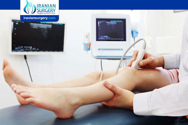
What happens during a Doppler ultrasound?
A sonographer, a specialist in ultrasound imaging technology, performs this test. The test may take 30 to 60 minutes.
Depending on the reason for the test, you may lie on your back or side on an exam table, or you may sit up.
During the test:
- The sonographer applies a small amount of gel to your skin. The gel helps the sonographer glide a small probe over the skin. It also helps sound waves travel.
- The transducer sends painless sound waves through your skin into your body. The sound waves are high frequency and you won’t hear them.
- The sound waves reflect off the moving blood cells, causing the pitch of the sound waves to change. You may hear a whooshing sound from the ultrasound machine.
- The transducer detects changes in the sound wave.
- A machine records the sound wave changes and converts them into images or graphs for your provider to review.
- The sonographer cleans the gel from your skin at the end of the test.
After Doppler Ultrasound
Results and Follow-up
When will I get the test results?
Your healthcare provider or a radiologist, a medical doctor who specializes in medical imaging, will review the test results. It may take a week to get the results.
Depending on the results, you may need more tests. The health condition will determine what additional tests you get.
Source:
. https://my.clevelandclinic.org/health/diagnostics/22715-doppler-ultrasound