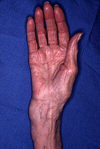
Distal radius fracture
A distal radius fracture, also known as wrist fracture, is a break of the part of the radius bone which is close to the wrist. Symptoms include pain, bruising, and rapid-onset swelling. The wrist may be deformed. The ulna bone may also be broken.
In younger people, these fractures typically occur during sports or a motor vehicle collision. In older people, the most common cause is falling on an outstretched hand. Specific types include Colles, Smith, Barton, and Chauffeur’s fractures. The diagnosis is generally suspected based on symptoms and confirmed with X-rays.
Treatment is with casting for six weeks or surgery. Surgery is generally indicated if the joint surface is broken and does not line up, the radius is overly short, or the joint surface of the radius is tilted more than 10% backwards. Among those who are cast, repeated X-rays are recommended within three weeks to verify that a good position is maintained.
Distal radius fractures are common. They represent between 25% and 50% of all broken bones. They occur most commonly in young males and older females. A year or two may be required for healing to occur.

Signs and symptoms
People usually present with a history of falling on an outstretched hand and complaint of pain and swelling around the wrist, sometimes with deformity around the wrist. Any numbness should be asked to exclude median and ulnar nerve injuries. Any pain in the limb of the same side should also be investigated to exclude associated injuries to the same limb.
Swelling, deformity, tenderness, and loss of wrist motion are normal features on examination of a person with a distal radius fracture. “Dinner fork” deformity of the wrist is caused by dorsal displacement of the carpal bones (Colle’s fracture). Reverse deformity is seen in volar angulation (Smith’s fracture). The wrist may be radially deviated due to shortening of the radius bone. Examination should also rule out a skin wound which might suggest an open fracture, usually at the side. Tenderness at an area with no obvious deformity may still point to underlying fractures. Decreased sensation over the thenar eminence can be due to median nerve injury. Swelling and displacement can cause compression on the median nerve which results in acute carpal tunnel syndrome and requires prompt treatment. Very rarely, pressure on the muscle components of the hand or forearm is sufficient to create a compartment syndrome.
Mechanism of injury
The most common cause of this type of fracture is a fall on an outstretched hand from standing height, although some fractures will be due to high-energy injury. People who fall on the outstretched hand are usually fitter and have better reflexes when compared to those with elbow or humerus fractures. The characteristics of distal radius fractures are influenced by the position of the hand at the time of impact, the type of surface at point of contact, the speed of the impact, and the strength of the bone. Distal radius fractures typically occur with the wrist bent back from 60 to 90 degrees. Radial styloid fracture would occur if the wrist is ulnar deviated and vice versa. If the wrist is bent back less, then proximal forearm fracture would occur, but if the bending back is more, then the carpal bones fracture would occur. With increased bending back, more force is required to produce a fracture. More force is required to produce a fracture in males than females. Risk of injury increases in those with osteoporosis.
Common injuries associated with distal radius fractures are interosseous intercarpal ligaments injuries, especially scapholunate (4.7% to 46% of cases) and lunotriquetral ligaments (12% to 34% of cases) injuries. There is an increased risk of interosseous intercarpal injury if the ulnar variance (the difference in height between the distal end of the ulna and the distal end of the radius) is more than 2mm and there is fracture into the wrist joint. Triangular fibrocartilage complex (TFCC) injury occurs in 39% to 82% of cases. Ulnar styloid process fracture increases the risk of TFCC injury by a factor of 5:1. However, it is unclear whether intercarpal ligaments and triangular fibrocartilage injuries are associated with long term pain and disability for those who are affected.
Diagnosis
Diagnosis may be evident clinically when the distal radius is deformed, but should be confirmed by X-ray. The differential diagnosis includes scaphoid fractures and wrist dislocations, which can also co-exist with a distal radius fracture. Occasionally, fractures may not be seen on X-rays immediately after the injury. Delayed X-rays, X-ray computed tomography (CT scan), or Magnetic resonance imaging (MRI) can confirm the diagnosis.
Medical imaging
X-ray of the affected wrist is required if a fracture is suspected. Posteroanterior, lateral, and oblique views can be used together to describe the fracture. X-ray of the uninjured wrist should also be taken to determine if any normal anatomic variations exist before surgery.
A CT scan is often performed to further investigate the articular anatomy of the fracture, especially for fracture and displacement within the distal radio-ulnar joint.
Various kinds of information can be obtained from X-rays of the wrist:
Lateral view
- Carpal malalignment – A line is drawn along the long axis of the capitate bone and another line is drawn along the long axis of the radius. If the carpal bones are aligned, both lines will intersect within the carpal bones. If the carpal bones are not aligned, both lines will intersect outside the carpal bones. Carpal malignment is frequently associated with dorsal or volar tilt of the radius and will have poor grip strength and poor forearm rotation.
- Tear drop angle – It is the angle between the line that pass through the central axis of the volar rim of the lunate facet of the radius and the line that pass through the long axis of the radius. Tear drop angle less than 45 degrees indicates displacement of lunate facet.
- Antero-posterior distance (AP distance) – Seen on lateral X-ray, it is the distance between the dorsal and volar rim of the lunate facet of the radius. The usual distance is 19 mm. Increased AP distance indicates the lunate facet fracture.
- Volar or dorsal tilt – A line is drawn joining the most distal ends of the volar and dorsal side of the radius. Another line perpendicular to the longitudinal axis of the radius is drawn. The angle between the two lines is the angle of volar or dorsal tilt of the wrist. Measurement of volar or dorsal tilt should be made in true lateral view of the wrist because pronation of the forearm reduces the volar tilt and supination increases it. When dorsal tilt is more than 11 degrees, it is associated with loss of grip strength and loss of wrist flexion.
Posteroanterior view
- Radial inclination – It is the angle between a line drawn from the radial styloid to the medial end of the articular surface of the radius and a line drawn perpendicular to the long axis of the radius. Loss of radial inclination is associated with loss of grip strength.
- Radial length – It is the vertical distance in milimetres between a line tangential to the articular surface of the ulna and a tangential line drawn at the most distal point of radius (radial styloid). Shortening of radial length more than 4mm is associated with wrist pain.
- Ulnar variance – It is the vertical distance between a horizontal line parallel to the articular surface of the radius and another horizontal line drawn parallel to the articular surface of the ulnar head. Positive ulnar variance (ulna appears longer than radius) disturbs the integrity of triangular fibrocartilage complex and is associated with loss of grip strength and wrist pain.
Oblique view
- Pronated oblique view of the distal radius helps to show the degree of comminution of the distal end radius, depression of the radial styloid, and confirming the position the screws at the radial side of the distal end radius. Meanwhile, a supinated oblique view of shows the ulnar side of the distal radius, accessing the depression of dorsal rim of the lunate facet, and the position of the screws on the ulnar side of the distal end radius.
Classification
There are many classification systems for distal radius fracture. AO/OTA classification is adopted by Orthopaedic Trauma Association and is the most commonly used classification system. There are three major groups: A—extra-articular, B—partial articular, and C—complete articular which can further subdivided into nine main groups and 27 subgroups depending on the degree of communication and direction of displacement. However, none of the classification systems demonstrate good liability. A qualification modifier (Q) is used for associated ulnar fracture.
Treatment
Correction should be undertaken if the wrist radiology falls outside the acceptable limits:
- 2-3mm positive ulnar varianceThere
- should be no carpus malalignment
- If carpus is aligned, then the dorsal tilt should be less than 10 degrees
- If carpus is aligned, there are no limits for palmar tilt
- If carpus is malaligned, wrist tilt should be neutral
- Gap or step deformity is less than 2mm
Treatment options for distal radius fractures include nonoperative management, external fixation, and internal fixation. Indications for each depend on a variety of factors such as the patient’s age, initial fracture displacement, and metaphyseal and articular alignment, with the ultimate goal to maximize strength and function in the affected upper extremity. Surgeons use these factors combined with radiologic imaging to predict fracture instability, and functional outcome to help decide which approach would be most appropriate. Treatment is often directed to restore normal anatomy to avoid the possibility of malunion, which may cause decreased strength in the hand and wrist. The decision to pursue a specific type of management varies greatly by geography, physician specialty (hand surgeons vs. orthopedic surgeons), and advancements in new technology such as the volar locking plating system.
Distal radius fractures are often associated with distal radial ulnar joint (DRUJ) injuries, and the American Academy of Orthopaedic Surgeons recommends that postreduction lateral wrist X-rays should be obtained in all patients with distal radius fractures in order to preclude DRUJ injuries or dislocations.
Reference:
https://en.wikipedia.org/wiki/Distal_radius_fracture
[kkstarratings]


