

Craniosynostosis treatment
Craniosynostosis
What is Craniosynostosis?
Craniosynostosis is a birth defect in which one or more of the fibrous joints between the bones of your baby's skull (cranial sutures) close prematurely (fuse), before your baby's brain is fully formed. Brain growth continues, giving the head a misshapen appearance.
Normally, during infancy the sutures remain flexible, giving your baby's brain time to grow. In the front of the skull, the sutures meet in the large soft spot (fontanel) on top of the head. The anterior fontanel is the soft spot you feel just behind your baby's forehead. The next largest is at the back (posterior). Each side of the skull has a tiny fontanel.
Craniosynostosis usually involves premature fusion of a single cranial suture, but can involve more than one of the sutures in your baby's skull (multiple suture craniosynostosis). In rare cases, craniosynostosis is caused by certain genetic syndromes (syndromic craniosynostosis).
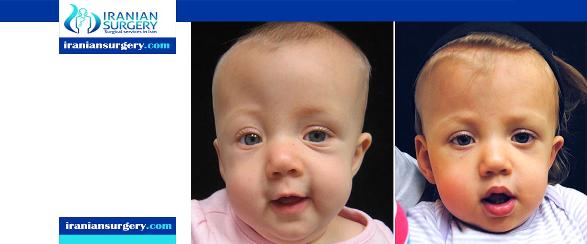
Treating craniosynostosis involves surgery to correct the shape of the head and allow for normal brain growth. Early diagnosis and treatment allow your baby's brain adequate space to grow and develop.
Although neurological damage can occur in severe cases, most children have normal cognitive development and achieve good cosmetic results after surgery. Early diagnosis and treatment are key.
Read more about : Cleft lip & Palate Repair
Before Craniosynostosis Treatment
Symptoms
The signs of craniosynostosis are usually noticeable at birth, but they'll become more apparent during the first few months of your baby's life. Signs and severity depend on how many sutures are fused and when in brain development the fusion occurs. These can include:
. A misshapen skull, with the shape depending on which of the sutures are affected
. An abnormal feeling or disappearing fontanel on your baby's skull
. Development of a raised, hard ridge along affected sutures
. Slow or no growth of the head as your baby grows
Read more about : Forehead reduction surgery
When to see a doctor
Your doctor will routinely monitor your child's head growth at well-child visits. Talk to your pediatrician if you have concerns about your baby's head growth or shape.
Causes
Often the cause of craniosynostosis is not known, but sometimes it's related to genetic disorders.
. Nonsyndromic craniosynostosis is the most common type of craniosynostosis, and its cause is unknown, although it's thought to be a combination of genes and environmental factors.
. Syndromic craniosynostosis is caused by certain genetic syndromes, such as Apert syndrome, Pfeiffer syndrome or Crouzon syndrome, which can affect your baby's skull development. These syndromes usually also include other physical features and health problems.
Read more about : Cochlear implants in Iran
Complications
What are the complications of craniosynostosis?
If untreated, craniosynostosis may cause, for example:
. Permanent head and facial deformity
. Poor self-esteem and social isolation
The risk of increased pressure inside the skull (intracranial pressure) from simple craniosynostosis is small, as long as the suture and head shape are fixed surgically. But babies with an underlying syndrome may develop increased intracranial pressure if their skulls don't expand enough to make room for their growing brains.
If untreated, increased intracranial pressure can cause:
. Developmental delays
. Cognitive impairment
. No energy or interest (lethargy)
. Blindness
. Eye movement disorders
. Seizures
. Death, in rare instances
Diagnosis
Craniosynostosis requires evaluation by specialists, such as a pediatric neurosurgeon or a specialist in plastic and reconstructive surgery. Diagnosis of craniosynostosis may include:
. Physical exam. Your doctor will feel your baby's head for abnormalities such as suture ridges, and look for facial deformities.
. Imaging studies. A computerized tomography (CT) scan or magnetic resonance imaging (MRI) of your baby's skull can show whether any sutures have fused. Cranial ultrasound imaging may be used. Fused sutures are identifiable by their absence, because they're invisible once fused, or by the ridging of the suture line. A laser scan and photographs also may be used to make precise measurements of the skull shape.
. Genetic testing. If your doctor suspects an underlying genetic syndrome, genetic testing may help identify the syndrome.
How can I prevent craniosynostosis?
There is no guaranteed way to prevent craniosynostosis. Prenatal genetic testing may show gene mutations that could lead to craniosynostosis. A genetic counselor can help you understand genetic risks and possible treatment options if your baby is born with craniosynostosis.
You can increase your chances of having a healthy baby by:
. Scheduling regular prenatal care visits.
. Speaking with your doctor about potential risk factors, including risks associated with fertility medications or thyroid disease.
. Taking prenatal vitamins or other supplements as directed.
How common is craniosynostosis?
Craniosynostosis is uncommon. It affects about 1 in every 2,500 babies in the United States.
Sagittal craniosynostosis is the most common type of congenital craniosynostosis.
During Craniosynostosis Treatment
Types of craniosynostosis
There are several types of craniosynostosis. Most involve the fusion of a single cranial suture. Some complex forms of craniosynostosis involve the fusion of multiple sutures. Most cases of multiple suture craniosynostosis are linked to genetic syndromes and are called syndromic craniosynostosis.
The term given to each type of craniosynostosis depends on what sutures are affected. Types of craniosynostosis include:
. Sagittal (scaphocephaly). Premature fusion of the sagittal suture that runs from the front to the back at the top of the skull forces the head to grow long and narrow. Sagittal craniosynostosis results in a head shape called scaphocephaly and is the most common type of craniosynostosis.
. Coronal. Premature fusion of one of the coronal sutures (unicoronal) that run from each ear to the top of the skull may cause the forehead to flatten on the affected side and bulge on the unaffected side. It also leads to turning of the nose and a raised eye socket on the affected side. When both coronal sutures fuse prematurely (bicoronal), the head has a short and wide appearance, often with the forehead tilted forward.
. Metopic. The metopic suture runs from the top of the bridge of the nose up through the midline of the forehead to the anterior fontanel and the sagittal suture. Premature fusion gives the forehead a triangular appearance and widens the back part of the head. This is also called trigonocephaly.
. Lambdoid. Lambdoid synostosis is a rare type of craniosynostosis that involves the lambdoid suture, which runs along the back of the head. It may cause one side of your baby's head to appear flat, one ear to be higher than the other ear and tilting of the top of the head to one side.
Treatment
Mild cases of craniosynostosis may not need treatment. Your doctor may recommend a specially molded helmet to help reshape your baby's head if the cranial sutures are open and the head shape is abnormal. In this situation, the molded helmet can assist your baby's brain growth and correct the shape of the skull.
However, for most babies, surgery is the primary treatment. The type and timing of surgery depends on the type of craniosynostosis and whether there's an underlying genetic syndrome. Sometimes more than one surgery is required.
The purpose of surgery is to correct the abnormal head shape, reduce or prevent pressure on the brain, create room for the brain to grow normally, and improve your baby's appearance. This involves a process of planning and surgery.
Surgical planning
Imaging studies can help surgeons develop a surgical procedure plan. Virtual surgical planning for treatment of craniosynostosis uses high-definition 3D CT scans and MRIs of your baby's skull to construct a computer-simulated, individualized surgical plan. Based on that virtual surgical plan, customized templates are constructed to guide the procedure.
Surgery
A team that includes a specialist in surgery of the head and face (craniofacial surgeon) and a specialist in brain surgery (neurosurgeon) generally performs the procedure. Surgery can be done by endoscopic or open surgery. Both types of procedures generally produce very good cosmetic results with low risk of complications.
. Endoscopic surgery. This minimally invasive surgery may be considered for babies up to age 6 months. Using a lighted tube and camera (endoscope) inserted through small scalp incisions, the surgeon removes the affected suture to enable your baby's brain to grow normally. Compared with an open procedure, endoscopic surgery has a smaller incision, typically involves only a one-night hospital stay and usually does not require a blood transfusion.
. Open surgery. Generally, for babies older than 6 months, open surgery is done. The surgeon makes an incision in the scalp and cranial bones, then reshapes the affected portion of the skull. The skull position is held in place with plates and screws that are absorbable. Open surgery typically involves a three- or four-day hospital stay, and blood transfusion is usually necessary. It's generally a one-time procedure, but in complex cases, multiple open surgeries are often required to correct the baby's head shape.
Helmet therapy
After endoscopic surgery, office visits at certain intervals are required to fit a series of helmets to help shape your baby's skull. The surgeon will determine the length of helmet therapy based on how quickly the shape responds to treatment. If open surgery is done, no helmet is needed afterward.
After Craniosynostosis Treatment
What is the outlook for babies with craniosynostosis?
Most babies who receive timely craniosynostosis treatment live a healthy life. Earlier treatment can minimize developmental problems due to pressure on the brain.
Sources:
. https://www.mayoclinic.org/diseases-conditions/craniosynostosis/symptoms-causes/syc-20354513
. https://my.clevelandclinic.org/health/articles/6000-craniosynostosis