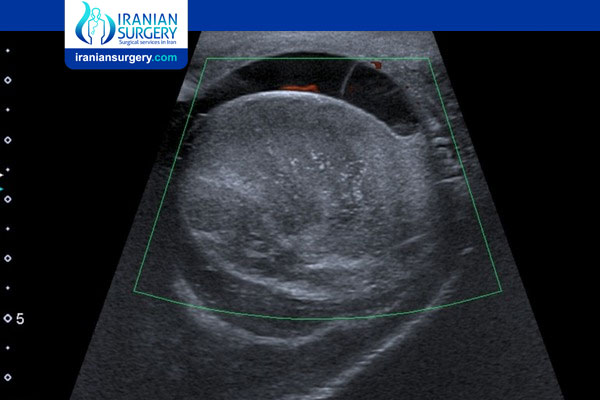
Testicular Ultrasound
Testicular Ultrasound
What is a Testicular Ultrasound?
A testicular ultrasound is a diagnostic test that obtains images of the testicles and the surrounding tissues in the scrotum. It’s also known as a testicular sonogram or scrotal ultrasound.
An ultrasound is a safe, painless, and noninvasive procedure in which high-frequency sound waves produce images of organs inside your body.
An ultrasound uses a probe or transducer. This handheld device converts energy from one form to another. An ultrasound technician, or sonographer, moves it against the targeted part of your body in sweeping motions.
The transducer emits sound waves as it moves. The transducer then receives the sound waves as they bounce off your organs in a series of echoes. A computer processes the echoes into images on a video monitor.
Normal and abnormal tissue transmit different types of echoes. A radiologist can interpret the echoes to distinguish between a benign condition and a solid mass that could be a malignant tumor.
Before Testicular Ultrasound
Why do I need a testicular ultrasound?
A testicular ultrasound is the primary imaging method used to observe and diagnose abnormalities in the testicles. A doctor may recommend a testicular ultrasound to:
. Determine the outcome of trauma to your scrotum
. Verify whether a lump in your scrotum or testicles is solid (which indicates a tumor) or filled with fluid (which indicates a cyst)
. Evaluate for possible testicular torsion, which is a twisted spermatic cord restricting blood flow to your testicle
. Identify sources of pain or swelling in your testicles
. Detect for and evaluate varicoceles, which are varicose veins within the scrotum
. Find the location of an undescended testicle
Ultrasound echoes can provide real-time still or moving images. Data from moving images is useful in examining blood flow to and from your testicles.
Are there risks involved with a testicular ultrasound?
A testicular ultrasound won’t put you at risk for any health problems. There’s no radiation exposure during the procedure.
However, you may have increased pain or discomfort during the procedure if you have certain testicular issues, such as testicular torsion or an infection.
How do I prepare for a testicular ultrasound?
Typically, there’s no special preparation necessary for a testicular ultrasound.
There’s no need to make dietary changes, fast, or maintain a full bladder before the exam. You won’t typically receive sedatives, anesthesia, or topical numbing agents.
There’s rarely a need to interrupt or discontinue medication use before a testicular ultrasound. However, you should still speak with your primary care physician about any prescription or over-the-counter (OTC) medications you take.
During Testicular Ultrasound
How is a testicular ultrasound performed?
A testicular ultrasound is usually an outpatient procedure performed in the radiology department of a hospital or at a doctor’s office.
A testicular ultrasound generally takes no more than 30 minutes. It involves the following steps.
. Positioning
You may need to change into a hospital gown.
Afterward, you’ll lie on your back with your legs spread. The ultrasound technician may place a towel underneath your scrotum to keep it elevated. They may place wide strips of tape across your thighs and under your scrotum to elevate your scrotum.
You’ll need to lie completely still during the procedure.
. Imaging technique
The technician will apply a warm, water-based gel to your testicles. This gel will allow the transducer to glide over your body. It also facilitates the conduction of the sound waves.
The technician will glide the transducer around your scrotum, moving back and forth. You may feel pressure as the technician pushes it firmly against your body. You may feel discomfort if there’s pressure on an area where you have tenderness due to an abnormality.
The technician will position the transducer against your body from different angles.
After Testicular Ultrasound
After the procedure
The technician will wipe the gel off your body after the procedure.
After your testicular ultrasound, you can resume your normal activities and diet. No recovery time is necessary.
What do the results mean?
A radiologist will analyze the images obtained during your testicular ultrasound. They’ll then send a report detailing the results of the test to your doctor.
Abnormal findings on a testicular ultrasound may indicate:
. An infection in your testicle
. Testicular torsion
. A testicular tumor
. A benign cyst
. A varicocele
. A hydrocele, which is a benign collection of fluid around your testicle
. A spermatocele, which is a fluid-filled cyst on the ducts of your testicle
Your doctor will probably recommend further investigation if the testicular ultrasound identifies a tumor.
Source:
. https://www.healthline.com/health/testicle-ultrasound


