

Microdochectomy
What is Microdochectomy
Microdochectomy is a procedure wherein a mammary or a lactiferous duct is removed. It is performed for patients that have nipple discharge coming from a single duct. It can be used for both diagnostic and therapeutic purposes.
Who Should Undergo and Expected Results
Microdochectomy, which can be both a diagnostic and therapeutic procedure, is recommended for patients experiencing nipple discharge. The condition is commonly caused by intraductal papilloma, which is responsible for approximately 80% of cases. This is a benign growth with an attachment to the wall of a mammary duct usually found just behind the nipple, and typically occurs in premenopausal women. An intraductal papilloma usually presents with either serous or bloody discharge coming from the nipple.
Nipple discharge can also be caused by:
- Some form of breast infection, such as mastitis or a breast abscess
- Galactorrhea and hormonal conditions, such as Cushing’s syndrome
- Duct ectasia, a benign change in the breast that is usually related to age
- Some medications, particularly contraceptive pills and some antidepressants, which may cause nipple discharge as a side effect
Although nipple discharge is usually associated with benign conditions, the risk of breast cancer in patients with this symptom increases if there is an associated mass and if the discharge is bloody. Based on statistics, 10% of patients with breast cancer also develop this symptom.

How is the Procedure Performed?
Microdochectomy is performed to manage nipple discharge that involves only a single duct. It is a simple outpatient procedure that can be carried out under either local or general anaesthesia. Prior to the surgery, the affected duct is identified through galactography, a procedure that investigates the ductal system of the breast and serves as a map of the ducts to locate the affected one. In addition, several examinations, including mammography and breast ultrasound, may be requested by the physician preoperatively.
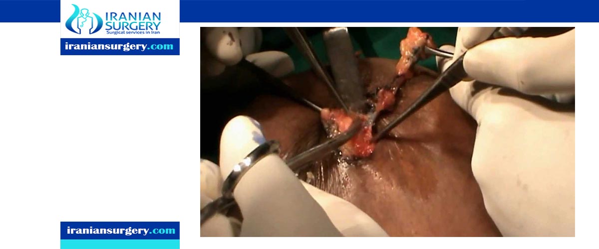
At the operating theatre, gentle pressure is exerted on the nipple to identify the orifice or opening of the affected duct. A fine probe is carefully inserted as far as possible into the duct, making sure that it is not damaged or disrupted. The duct is then dilated and dye is injected into it, marking the duct.
The borders of the nipple are then traced and incised (circumareolar incision). The areolar skin is raised up to create a skin flap. The affected duct is gently dissected and freed off from the tissues surrounding it for approximately 5 cm. The duct is then transected and removed. Some surgeons may opt to insert a drain, which will be removed after several hours. Closure of the incision is accomplished using absorbable sutures.
Microdochectomy serves as both a diagnostic and a therapeutic procedure. The specimen collected is sent for biopsy to determine the cause of the nipple discharge. If only a single duct is involved, microdochectomy will result in the resolution of the nipple discharge. However, if multiple ducts are involved, a more extensive procedure, such as a subareolar resection or central duct excision, may be indicated.
The major benefit of performing a microdochectomy is the preservation of the patient’s ability to breastfeed. This advantage is ideal for young patients who are currently breastfeeding or who have plans of breastfeeding in the future.
Possible Risks and Complications
Microdochectomy is relatively straightforward with minimal complications. A frequent problem encountered when performing the procedure is difficulty identifying the affected duct. In these cases, one option that can be done is the exploration of the subareolar region.
Meanwhile, the usual complications of the procedure involve the wound, which include:
- Infections, which can occur especially in patients with recurrent infections of the breast. This can be managed with proper antibiotic treatment.
- Pain, which is usually tolerable and may be managed with pain medications.
- Bruising and hematoma formation, which are uncommon complications, but can also occur after the procedure.
- Poor cosmetic outcomes
- Poor wound healing, which can result in permanent changes in the shape and colour of the nipple, resulting in asymmetry and a dark scar (hyperpigmentation). In some cases, poor healing in deep locations can result in a retracted nipple or a palpable lump in the breast after the operation.
Other possible complications include:
- Change in nipple sensation - The nerves supplying the nipple may be accidentally transected or stretched, resulting in numbness, or occasionally, pain.
- Losing the ability to breastfeed - Although microdochectomy allows the preservation of breastfeeding ability, there have been a few reports stating loss of this ability following the procedure.
[kkstarratings]