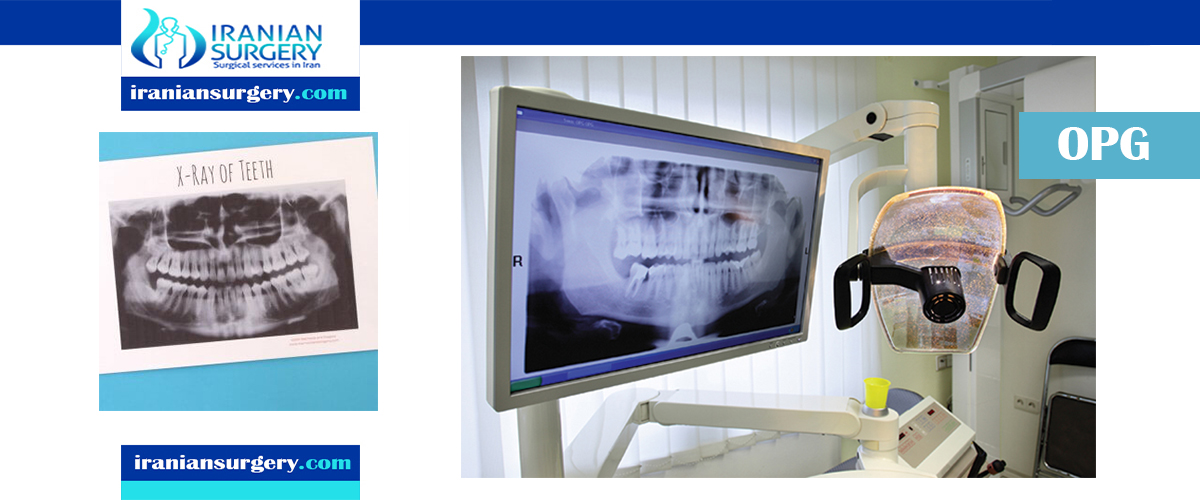
Dental X-ray (OPG)

dental opg iran

What Is Orthopantomography?

Orthopantomography, also known as an OPG X-ray (or simply OPG), panoramic radiography, or a pantogram is a type of X-ray scan that gives a panoramic or wide view of the lower face. It can display all the teeth on both jaws on one film, including those that have not surfaced or erupted yet, such as wisdom teeth. It also offers a view of the jawbone and the temporomandibular joint (TMJ), which connects the jaw to the rest of the skull.

Read more about : Dental implant in Iran

What Does An OPG X-Ray Involve?

An OPG is an X-ray of the lower face. Like all X-rays, it involves using short blasts of low-level radiation to create images of inside the body – in this case, of the bones and teeth.

The procedure for dental panoramic radiography consists of the patient resting their chin on a small shelf in front of the X-ray machine and bite softly on a sterile mouthpiece. This will keep the head and mouth steady while the images are taken.

The panoramic X-ray machine consists of a rotating arm with the X-ray source at one end and the film mechanism (which captures the image) at the opposite extremity. The arm rotates around the patient’s head to capture the wide view of their mouth and jaw.

The procedure is performed very quickly. As with any X-ray, the patient feels no discomfort during the procedure and can continue with their daily routine afterwards.

Read more about : Root canal trearment

Why Is an OPG X-Ray Done?

Orthopantomography is a technique used in dentistry to allow the dentist to view all their patient’s teeth and determine their number, position, and growth, including those that have not yet erupted. An OPG X-ray might be done to plan orthodontic treatment, to detect the presence or asses the development of wisdom teeth, to examine the jawbone, or for a general overview of the patient’s dental health.

What happens during a dental x-ray?

You may be asked to remove jewelry, eyeglasses, and any metal objects that may obscure the images. You will be asked to stand with your face resting on a small shelf and to bite gently on a sterile mouth piece to steady your head. It is important to stay very still while the x-ray is taken. You will not feel any discomfort during the procedure.

OPG Advances / developments

Dental X-ray technology is currently moving away from traditional film technology to digital X-ray technology, using electronic sensors and computers to create images. Digital X-rays allow instant review of the scans without having to wait to develop the film. They are also more efficient at getting high-quality images first time, reducing the number of repeat scans necessary, and therefore reducing the patient’s exposure to radiation.

Source:

https://www.topdoctors.co.uk/medical-dictionary/opg-x-ray-orthopantomography

10 common question about dental opg

1What is OPG in dental?
Dental X-ray (OPG) An OPG (Orthopantomogram) is a scan that gives a panoramic view of your jaw and teeth. The scan can provide information on wisdom teeth, bone loss, orthodontic assessment, jaw trauma, dental pain, or be used as part of a general dental check-up.
2How many dental xrays are safe?
When these x-rays are performed properly with adequate safety precautions in place, there's very little cause for concern. A routine examination with four bitewing x-rays exposes you to roughly the same amount of radiation you will experience during one to two hours on an airplane.
3How is OPG done?
An OPG is a panoramic X-ray of the upper and lower jaws, including the teeth. The OPG unit is specifically designed to rotate around the patient's head during the scan. An OPG will take approximately 20 seconds. It can also be used for surgical planning.
4How is OPG taken?
During OPG x-rays, you will be asked to stand or sit in front of the x-ray machine and rest your chin on a plastic rest. You will also need to bite down gently on a mouthpiece, to ensure your head remains still during the procedure. Part of the machine then rotates around your head as the images are being taken.
5Are teeth xrays dangerous?
Risks of dental X-rays While dental X-rays do involve radiation, the exposed levels are so low that they're considered safe for children and adults. If your dentist uses digital X-rays instead of developing them on film, your risks from radiation exposure are even lower.
6Can dental Xrays cause cancer?
April 10, 2012 -- Getting frequent dental X-rays appears to increase the risk for a commonly diagnosed brain tumor, a new study finds. Exposure to ionizing radiation -- the kind found in X-rays -- is the biggest known environmental risk factor for largely non-malignant meningioma brain tumors.
7Are xrays dangerous?
Generally, the amount of radiation you're exposed to during an X-ray is the equivalent to between a few days and a few years of exposure to natural radiation from the environment. Being exposed to X-rays does carry a risk of causing cancer many years or decades later, but this risk is thought to be very small.
8What is OPG bone?
Osteoprotegerin (OPG) is secreted by osteoblasts and osteogenic stromal stem cells and protects the skeleton from excessive bone resorption by binding to RANKL and preventing it from interacting with RANK. The RANKL/OPG ratio in bone marrow is thus an important determinant of bone mass in normal and disease states.
9What does Cbct stand for?
Cone beam computed tomography Cone beam computed tomography (or CBCT, also referred to as C-arm CT, cone beam volume CT, or flat panel CT) is a medical imaging technique consisting of X-ray computed tomography where the X-rays are divergent, forming a cone.
10What is cephalometric radiograph?
A Cephalometric radiograph is a radiograph of the head taken in a Cephalometer (Cephalostat) that is a head-holding device introduced in 1931 by Holly Broadbent Sr. in USA. The Cephalometer is used to obtain standardized and comparable craniofacial images on radiographic films.

Leave a Reply

Your email address will not be published. Required fields are marked *

Patient Review