

Corneal opacity treatment
Types Of Corneal Opacity
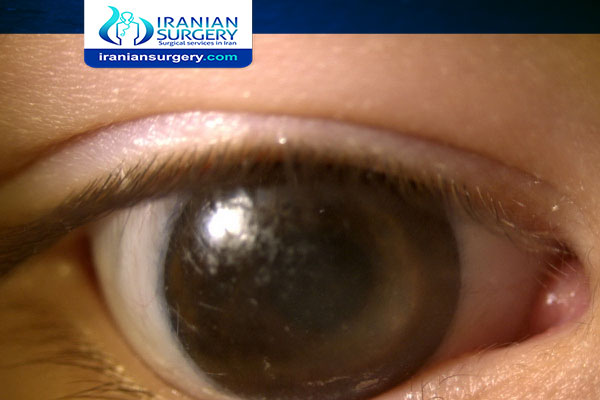
What Is Corneal Opacity?
Corneal opacities are eye problems that can lead to scarring or clouding of the cornea, which decreases vision. The cornea is the clear, dome-shaped area that covers the front of the eye. Light passes through the cornea before reaching the retina in the back of the eye, and so it must remain clear so light can pass through.
Corneal opacities can cause anything from minor irritation to vision problems and even blindness. In fact, corneal problems are the fourth leading cause of blindness (after glaucoma, cataracts, and age-related macular degeneration). Injury, infection, and certain eye diseases can cause corneal opacities.
Types Of Corneal Opacity
Depending on the density, corneal opacity is graded as nebula, macula and leucoma.
Nebular Corneal Opacity
It is a faint opacity which results due to superficial scars involving Bowman's layer and superficial stroma. A nebular corneal opacity allows the details of the iris to be seen through the opacity. A thin, diffuse nebula covering the pupillary area interferes more with vision than a strictly localized dense leucoma, so long as the latter does not block the whole pupillary area. This is because the leucoma stops all the light which falls upon it, whereas the nebula refracts it irregularly, allowing many of the rays to fall upon the retina where they blur the image formed by the regularly refracted rays.
Macular Corneal Opacity
It is a semi-dense opacity produced when scarring involves about half the corneal stroma.
Leucomatous Corneal Opacity (Leucoma Simplex)
It is a dense white opacity which results due to scarring of more than half of the stroma
. Adherent leucoma: It results when healing occurs after perforation of cornea with incarceration of iris. The iris is adherent to the back of a leucomatous cornea. One of the major complications of adherent leucoma is Secondary glaucoma
. Corneoiridic scar: If iris tissue is incarcerated and incorporated within the scar tissue, as occurs in healing of a large sloughed corneal ulcer, it is called a corneoiridic scar.
. Corneal facet: Sometimes the corneal surface is depressed at the site of healing (due to less fibrous tissue); such a scar is called facet.
. Kerectasia: In this condition, corneal curvature is increased at the site of opacity (bulge due to weak scar).
About Iranian Surgery
Iranian surgery is an online medical tourism platform where you can find the best ophthalmologists in Iran. The price of Corneal Opacity Treatment in Iran can vary according to each individual’s case and will be determined by an in-person assessment with the doctor.
For more information about the cost of Corneal Opacity Treatment in Iran and to schedule an appointment in advance, you can contact Iranian Surgery consultants via WhatsApp number 0098 901 929 0946. This service is completely free.
Source:
2 Comments
is it true that one of the causes of corneal opacity is glaucoma???
Congenital corneal opacities are mostly stemmed from a malformation of the anterior segment of the eye (anterior segment dysgenesis). However, there are additional causes including congenital glaucoma, dermoid, trauma, infection, corneal dystrophies, and metabolic storage diseases.