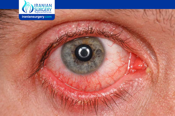
Retinal detachment
Retinal Detachment
What is Retinal Detachment?
Retinal detachment describes an emergency situation in which a thin layer of tissue (the retina) at the back of the eye pulls away from its normal position.
Retinal detachment separates the retinal cells from the layer of blood vessels that provides oxygen and nourishment. The longer retinal detachment goes untreated, the greater your risk of permanent vision loss in the affected eye.
Warning signs of retinal detachment may include one or all of the following: the sudden appearance of floaters and flashes and reduced vision. Contacting an eye specialist (ophthalmologist) right away can help save your vision.
About Iranian Surgery
Iranian surgery is an online medical tourism platform where you can find the best ophthalmologists in Iran. The price of Retinal Detachment treatment in Iran can vary according to each individual’s case and will be determined by an in-person assessment with the doctor.
For more information about the cost of Retinal Detachment Treatment in Iran and to schedule an appointment in advance, you can contact Iranian Surgery consultants via WhatsApp number 0098 901 929 0946. This service is completely free.
Before Retinal Detachment Treatment
Symptoms
Retinal detachment itself is painless. But warning signs almost always appear before it occurs or has advanced, such as:
. The sudden appearance of many floaters — tiny specks that seem to drift through your field of vision
. Flashes of light in one or both eyes (photopsia)
. Blurred vision
. Gradually reduced side (peripheral) vision
. A curtain-like shadow over your visual field
When to see a doctor
Seek immediate medical attention if you are experiencing the signs or symptoms of retinal detachment. Retinal detachment is a medical emergency in which you can permanently lose your vision.
Causes
There are three different types of retinal detachment:
. Rhegmatogenous (reg-ma-TODGE-uh-nus). These types of retinal detachments are the most common. Rhegmatogenous detachments are caused by a hole or tear in the retina that allows fluid to pass through and collect underneath the retina, pulling the retina away from underlying tissues. The areas where the retina detaches lose their blood supply and stop working, causing you to lose vision.
The most common cause of rhegmatogenous detachment is aging. As you age, the gel-like material that fills the inside of your eye, known as the vitreous (VIT-ree-us), may change in consistency and shrink or become more liquid. Normally, the vitreous separates from the surface of the retina without any complications — a common condition called posterior vitreous detachment (PVD). One complication of this separation is a tear.
As the vitreous separates or peels off the retina, it may tug on the retina with enough force to create a retinal tear. Left untreated, the liquid vitreous can pass through the tear into the space behind the retina, causing the retina to become detached.
. Tractional. This type of detachment can occur when scar tissue grows on the retina's surface, causing the retina to pull away from the back of the eye. Tractional detachment is typically seen in people who have poorly controlled diabetes or other conditions.
. Exudative. In this type of detachment, fluid accumulates beneath the retina, but there are no holes or tears in the retina. Exudative detachment can be caused by age-related macular degeneration, injury to the eye, tumors or inflammatory disorders.
Risk factors
The following factors increase your risk of retinal detachment:
. Aging — retinal detachment is more common in people over age 50
. Previous retinal detachment in one eye
. Family history of retinal detachment
. Extreme nearsightedness (myopia)
. Previous eye surgery, such as cataract removal
. Previous severe eye injury
. Previous other eye disease or disorder, including retinoschisis, uveitis or thinning of the peripheral retina (lattice degeneration)
Diagnosis
Your doctor may use the following tests, instruments and procedures to diagnose retinal detachment:
. Retinal examination. The doctor may use an instrument with a bright light and special lenses to examine the back of your eye, including the retina. This type of device provides a highly detailed view of your whole eye, allowing the doctor to see any retinal holes, tears or detachments.
. Ultrasound imaging. Your doctor may use this test if bleeding has occurred in the eye, making it difficult to see your retina.
Your doctor will likely examine both eyes even if you have symptoms in just one. If a tear is not identified at this visit, your doctor may ask you to return within a few weeks to confirm that your eye has not developed a delayed tear as a result of the same vitreous separation. Also, if you experience new symptoms, it's important to return to your doctor right away.
During Retinal Detachment Treatment
Treatment
Surgery is almost always used to repair a retinal tear, hole or detachment. Various techniques are available. Ask your ophthalmologist about the risks and benefits of your treatment options. Together you can determine what procedure or combination of procedures is best for you.
Retinal tears
When a retinal tear or hole hasn't yet progressed to detachment, your eye surgeon may suggest one of the following procedures to prevent retinal detachment and preserve vision.
. Laser surgery (photocoagulation). The surgeon directs a laser beam into the eye through the pupil. The laser makes burns around the retinal tear, creating scarring that usually "welds" the retina to underlying tissue.
. Freezing (cryopexy). After giving you a local anesthetic to numb your eye, the surgeon applies a freezing probe to the outer surface of the eye directly over the tear. The freezing causes a scar that helps secure the retina to the eye wall.
Both of these procedures are done on an outpatient basis. After your procedure, you'll likely be advised to avoid activities that might jar the eyes — such as running — for a couple of weeks or so.
Retinal detachment
If your retina has detached, you'll need surgery to repair it, preferably within days of a diagnosis. The type of surgery your surgeon recommends will depend on several factors, including how severe the detachment is.
. Injecting air or gas into your eye. In this procedure, called pneumatic retinopexy (RET-ih-no-pek-see), the surgeon injects a bubble of air or gas into the center part of the eye (the vitreous cavity). If positioned properly, the bubble pushes the area of the retina containing the hole or holes against the wall of the eye, stopping the flow of fluid into the space behind the retina. Your doctor also uses cryopexy during the procedure to repair the retinal break.
Fluid that had collected under the retina is absorbed by itself, and the retina can then adhere to the wall of your eye. You may need to hold your head in a certain position for up to several days to keep the bubble in the proper position. The bubble eventually will reabsorb on its own.
. Indenting the surface of your eye. This procedure, called scleral (SKLAIR-ul) buckling, involves the surgeon sewing (suturing) a piece of silicone material to the white of your eye (sclera) over the affected area. This procedure indents the wall of the eye and relieves some of the force caused by the vitreous tugging on the retina.
If you have several tears or holes or an extensive detachment, your surgeon may create a scleral buckle that encircles your entire eye like a belt. The buckle is placed in a way that doesn't block your vision, and it usually remains in place permanently.
. Draining and replacing the fluid in the eye. In this procedure, called vitrectomy (vih-TREK-tuh-me), the surgeon removes the vitreous along with any tissue that is tugging on the retina. Air, gas or silicone oil is then injected into the vitreous space to help flatten the retina.
Eventually the air, gas or liquid will be absorbed, and the vitreous space will refill with body fluid. If silicone oil was used, it may be surgically removed months later.
Vitrectomy may be combined with a scleral buckling procedure.
After surgery your vision may take several months to improve. You may need a second surgery for successful treatment. Some people never recover all of their lost vision.
After Retinal Detachment Treatment
Recovery
You have had surgery to fix a retinal detachment. Your doctor may also have fixed a tear in your retina.
Your eye doctor may put drops in your eye to prevent infection and keep the pupil from opening wide or closing. You will also use these drops at home. You may have to wear a patch or shield over the eye for a day or more. You may have some pain in your eye and your vision may be blurry for a few days after the surgery. Your eye may be swollen, red, or tender for several weeks.
If your doctor used a gas bubble to flatten your retina during surgery, you may have to keep your head in a special position for a few days or longer. Your doctor will give you special instructions about this.
You will need 2 to 4 weeks to recover before returning to your normal activities.
This care sheet gives you a general idea about how long it will take for you to recover. But each person recovers at a different pace. Follow the steps below to get better as quickly as possible.
How can you care for yourself at home?
. Activity
. Rest when you feel tired.
. Allow the eye to heal. Don't do things where you might move your head. This includes moving quickly, lifting anything heavy, or doing activities such as cleaning or gardening.
. You will probably need to take 2 to 4 weeks off from work. It depends on the type of work you do and how you feel.
. You may drive when your vision allows it. If you are not sure, ask your doctor.
. Diet
. You can eat your normal diet. If your stomach is upset, try bland, low-fat foods like plain rice, broiled chicken, toast, and yogurt.
. Medicines
. Your doctor will tell you if and when you can restart your medicines. You will also get instructions about taking any new medicines.
. If you take aspirin or some other blood thinner, ask your doctor if and when to start taking it again. Make sure that you understand exactly what your doctor wants you to do.
. Be safe with medicines. Read and follow all instructions on the label.
. If the doctor gave you a prescription medicine for pain, take it as prescribed.
. If you are not taking a prescription pain medicine, ask your doctor if you can take an over-the-counter medicine.
. You will need to use eyedrops for up to 6 weeks.
. Ice and elevation
. Close your eye and put ice or a cold pack on it for 10 to 20 minutes at a time. Try to do this every 1 to 2 hours for the next 3 days (when you are awake) or until the swelling goes down. Put a thin cloth between the ice and your skin.
. Other instructions
. You can shower and wash your hair and face. But don't get any soap in your eye. You may want to use a face cloth when you wash your face. Some people wear swimming goggles.
. Wear sunglasses during the day. You may have to wear an eye patch or shield for a few days.
. If your doctor used a gas bubble to hold the retina in place, keep your head in a certain position for most of the day and night for 1 to 3 weeks after the surgery. Your doctor will give you specific instructions.
. Do not lie on your back. The bubble will move to the front of the eye and press against the lens instead of the retina.
. Airplane travel is dangerous. This is because the change in altitude may cause the gas bubble to expand and increase the pressure inside the eye.
Follow-up care is a key part of your treatment and safety. Be sure to make and go to all appointments, and call your doctor or nurse if you are having problems. It's also a good idea to know your test results and keep a list of the medicines you take.
When should you call for help?
Call your doctor or nurse call line now or seek immediate medical care if:
. You have sudden chest pain, shortness of breath, or you cough up blood.
. You have symptoms of an eye infection, such as:
. Pus or thick discharge coming from the eye.
. Redness or swelling around the eye.
. A fever.
. You have new or worse eye pain.
. You have vision changes.
. You see new flashes of light.
. You have symptoms of a blood clot in your leg (called a deep vein thrombosis), such as:
. Pain in the calf, back of the knee, thigh, or groin.
. Redness and swelling in your leg or groin.
Watch closely for changes in your health, and be sure to contact your doctor or nurse if:
. You see new floaters.
. You do not get better as expected.
Sources:
. https://www.mayoclinic.org/diseases-conditions/retinal-detachment/symptoms-causes/syc-20351344
.https://myhealth.alberta.ca/Health/aftercareinformation/pages/conditions.aspx?hwid=abp2623
10 common question about retinal detachment
[kkstarratings]


