

peripheral angioplasty with vascular closure device
peripheral angioplasty with vascular closure device (vcd)
A vascular closure device is usually a piece of collagen (a fibrous protein found in skin, bone and connective tissue), metallic clip or suture (stitch) designed to provide immediate sealing of the small puncture made in an artery after an angiogram.
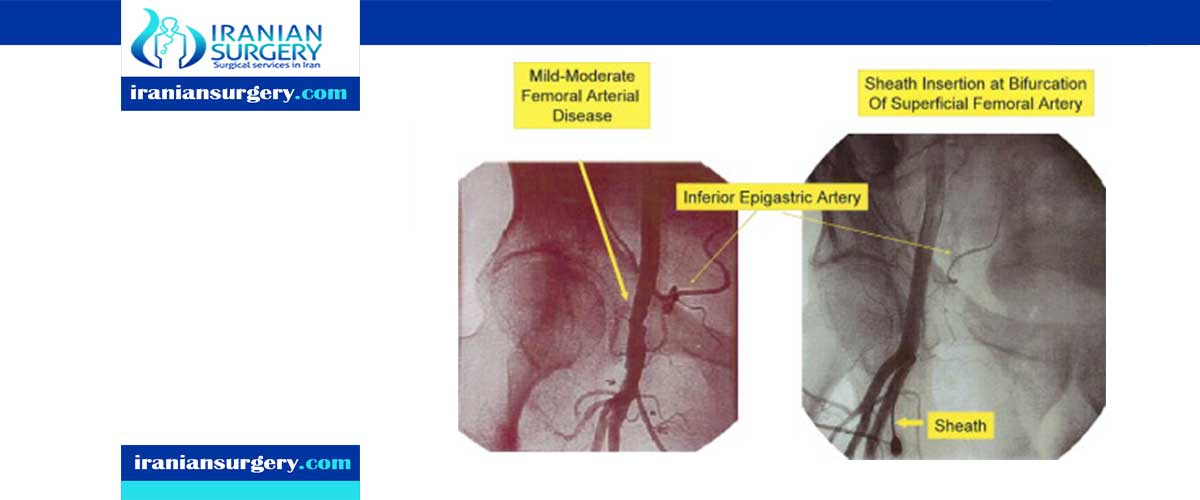
Angiography is the imaging of blood vessels that appear on live X-ray images or pictures. This helps show abnormalities or to guide the treatment of disease. Arterial access is commonly carried out through the femoral artery, which is a large artery running past the region of the groin. Access through this artery involves a specialist doctor puncturing the artery with a small needle through the skin and inserting a plastic sheath or tube into the artery through the puncture site.

Once the angiographic procedure is complete, the plastic sheath providing access to the femoral artery will need to be removed. This will leave behind a small hole in the artery, corresponding to the size of the plastic sheath. The hole will continue to bleed, unless treated. Stopping the flow of blood from this hole is normally achieved by manual compression with the doctor’s or nurse’s fingers placed directly over the site of the hole in the artery.
Manual compression is usually very effective, but can take 10–15 minutes or longer, depending on the size of the plastic sheath placed in the artery. Manual compression requires patients to lie flat in bed with the hips in a straight position for at least 4 hours after the plastic sheath is removed.
Vascular closure devices provide an alternative to manual compression. These devices provide immediate sealing of the femoral artery access site, so there is no need for prolonged compression.
10 common questions about peripheral angioplasty with vascular closure device
[kkstarratings]